Recombinant Human PD-1 (CD279)-Fc Chimera (carrier-free) 10 µg

Produit ni repris ni échangé excepté en cas d’erreur du prestataire.
Points clés
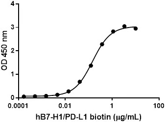
Programmed death-1 (PD-1) is a type I transmembrane protein initially isolated from apoptosis-induced cells by subtractive hybridization. The amino acid sequence of mouse PD-1 shares 59.4% identity to the human counterpart, and a putative tyrosine kinase-association motif is well conserved. Its extracellular region consists of a single Ig-like variable (IgV) domain. PD-1 is an inhibitory molecule expressed by activated B and T cells and has been implicated in immune tolerance. PD-L1 and PD-L2 are the ligands for PD-1 and their binding leads to the inhibition of T cell receptor-mediated lymphocyte proliferation and cytokine secretion. PD-1/PD-L1 interaction suppresses immune responses against autoantigens and tumors, and plays an important role in the maintenance of peripheral immune tolerance. Interaction between PD-1 and PD-L1 promote tolerance by blocking TCR-mediated signaling. PD-L1 expression by tumor cells allow tumor progression by suppressing antitumor T-cell responses. Antibodies against PD-1 or PD-L1 lead to increased antitumor immunity.;
Garantie
Garantie 0 Mois
Description
Programmed death-1 (PD-1) is a type I transmembrane protein initially isolated from apoptosis-induced cells by subtractive hybridization. The amino acid sequence of mouse PD-1 shares 59.4% identity to the human counterpart, and a putative tyrosine kinase-association motif is well conserved. Its extracellular region consists of a single Ig-like variable (IgV) domain. PD-1 is an inhibitory molecule expressed by activated B and T cells and has been implicated in immune tolerance. PD-L1 and PD-L2 are the ligands for PD-1 and their binding leads to the inhibition of T cell receptor-mediated lymphocyte proliferation and cytokine secretion. PD-1/PD-L1 interaction suppresses immune responses against autoantigens and tumors, and plays an important role in the maintenance of peripheral immune tolerance. Interaction between PD-1 and PD-L1 promote tolerance by blocking TCR-mediated signaling. PD-L1 expression by tumor cells allow tumor progression by suppressing antitumor T-cell responses. Antibodies against PD-1 or PD-L1 lead to increased antitumor immunity.;
Caractéristiques
- Fournisseur
- BioLegend Europe BV
- Marque
- BIOLEGEND
- Référence fabricant
- 785102
- Référence distributeur
- 785102
- Vendu par
- 10 μg
- Quantité
- N/A
- Lieu de fabrication
- USA
- Lieu de stockage
- Pays-Bas ou USA
- Référence fabriquant similaire
- 785106
- Soumis à carboglace
- non
- Classement dans le catalogue fournisseur
- Recombinant Protein
- Certification
- RUO
- Type d’application
- bioassay
- Type de produit
- Recombinant Protein
- Température de conservation (°C)
- -20 ou -70 °C
- Température de transport
- Blue Ice
- Organisme cible
- Human
- Source biologique
- 293E cells
- Seuil de coupure des masses moléculaires MWCO
- The 382 amino acid recombinant protein has a predicted molecular mass of approximately 42.8 kD. The DTT-reduced protein migrates at approximately 60 - 70 kD and and non-reduced protein migrates at approximately 100 - 120 kD by SDS-PAGE. Da
- Concentration
- 10 and 25 µg sizes are bottled at 200 µg/mL. 100 µg and larger sizes are lot-specific and bottled at the concentration indicated on the vial. To obtain lot-specific concentration, please enter the lot number in our online tools.
- Pureté
- >95%, as determined by Coomassie stained SDS-PAGE. %
- Matière dangereuse
- Non
- Code douanier
- 38220000
- Classement NCBI
- 5133
- Nomenclature Nacres
- NA.77
- Nomenclature CEA
- SGP01
- Nomenclature IRSN
- 273
- Nomenclature INSERM
- NA.NA77
- Nomenclature CNRS
- NA77
- Nomenclature CHU
- 18.551
- Nomenclature DGOS
- LD11AOOO
- Reprise en cas d’erreur client
- non
